Retinal Detachment: Prevention and Management

Retinal Detachment: Prevention and Management

Retinal Detachment: Prevention and Management

Retinal Detachment: Prevention and Management

Retinal Detachment: Prevention and Management

Retinal Detachment: Prevention and Management

What Is Retinal Detachment
When it becomes detached, this communication is disrupted, potentially leading to partial or complete vision loss if left untreated.
This condition can develop suddenly or gradually, and while it is most common in older adults, it can affect individuals across various age groups. The detachment itself can occur due to tears or breaks in the retina that allow fluid to collect underneath. This separation deprives retinal cells of nourishment and oxygen, which can cause irreversible damage if not promptly addressed.
Exudative Retinal DetachmentIt may result from inflammation, injury, or certain systemic conditions.
Each type presents unique challenges for diagnosis and treatment but shares one key characteristic: all require immediate attention by an ophthalmologist for an optimal outcome.
Understanding what retinal detachment entails highlights its gravity as a medical emergency. Even though modern advancements offer effective treatments like laser surgery and vitrectomy for many cases, prevention remains crucial since vision loss from prolonged detachment can often be permanent. Recognizing symptoms early on and seeking timely care are essential in preserving long-term eye health.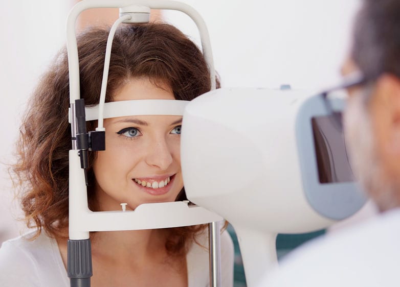
Common Causes and Risk Factors
Retinal detachment is a serious eye condition that can develop from various underlying causes, many of which are preventable with proper care and awareness. Understanding the common causes and risk factors associated with this condition is crucial for early intervention and effective management. This separation disrupts vision and, if left untreated, can lead to permanent blindness.
One of the most prevalent causes of retinal detachment is posterior vitreous detachment (PVD), a condition in which the gel-like substance inside the eye (the vitreous) shrinks and pulls away from the retina. While PVD is a normal part of aging for many people, in some cases it creates traction on the retina, leading to tears or holes that may progress to detachment.
Blunt force trauma can lead to retinal tears or detachments by creating direct damage to retinal tissue or increasing intraocular pressure, which destabilizes its delicate structure. Athletes involved in high-contact sports like boxing or soccer should take precautions to protect their eyes using protective eyewear.
Genetics also plays a prominent role in determining risk levels. If you have a family history of retinal detachment, particularly among first-degree relatives such as parents or siblings, your susceptibility may be higher than average. Additionally, individuals who are highly myopic (nearsighted) often have longer eyeballs that can stretch and thin parts of their retinas over time, making them more vulnerable.
Certain medical conditions increase risks as well. People with diabetes may develop diabetic retinopathy—a condition where abnormal blood vessels grow on the retina’s surface—heightening their chances of tears or detachment.
Inflammatory disorders affecting ocular tissues such as uveitis can also weaken attachments within the eye's internal structures over time, increasing vulnerability. Lastly, lifestyle-related factors like smoking are believed by researchers to contribute indirectly by damaging blood vessels throughout the body—including those supplying oxygen-rich blood critical for retinal function.
Recognizing these causes allows individuals at risk to take proactive measures such as regular eye exams and adopting healthy habits that promote overall ocular wellness. While some genetic predispositions cannot be mitigated entirely through behavior changes alone, addressing modifiable risks—including controlling blood sugar levels if diabetic or wearing appropriate protective gear during physical activities—can go a long way toward safeguarding eyesight against this debilitating condition.
Early Warning Signs You Should Never Ignore
Recognizing the early warning signs of retinal detachment can be the difference between preserving your vision and permanent vision loss. Retinal detachment typically begins with subtle symptoms, but these can escalate quickly, making it critical to act immediately if they occur. Early detection followed by prompt medical intervention offers the best chance of preventing severe complications.
One of the most commonly reported early signs is the sudden appearance of floaters. These are small, shadowy shapes that may look like specks, strings, or cobwebs drifting in your field of vision. While floaters are common with aging and not always cause for alarm, a sudden increase in their number or size could signal a problem with the retina.
Another significant symptom is flashes of light, which may appear in your peripheral vision or even when your eyes are closed. These flashes occur when the retina is tugged by vitreous gel within the eye. This sensation often feels like brief sparks or streaks of light and could indicate that the retina is under stress or beginning to detach.
A third critical sign to watch for is a shadow or curtain effect over your visual field. This may start in one corner and progressively expand across your sight as more of the retina becomes detached from its underlying support structure. This symptom often points to full-thickness detachment and requires immediate evaluation by an eye care professional.
Blurred or distorted vision can also serve as an early indicator. If fine details suddenly become harder to focus on or straight lines begin appearing wavy or warped, it could mean that fluid has begun accumulating under the retina or that it’s shifted out of its proper position.
Keep in mind that retinal detachment itself is usually painless due to the lack of pain receptors in the retina. However, ignoring these visual disturbances can lead to severe and irreversible damage because once cells within the detached area stop receiving oxygen and nutrients from nearby blood vessels, they begin to die off rapidly.
If you experience any combination of these symptoms—particularly if they occur suddenly—it’s important to contact an ophthalmologist right away for an emergency evaluation. Timely treatment can prevent further progression and potentially save your sight altogether.
While it’s essential to recognize these signs yourself, regular eye exams play a vital role in identifying underlying risk factors before symptoms even arise. Staying informed about what constitutes normal versus abnormal changes in vision empowers you to take charge of your eye health proactively.
How Retinal Detachment Is Diagnosed
Diagnosing retinal detachment promptly is crucial for preserving vision and preventing further complications. Diagnostic procedures involve a detailed evaluation by an ophthalmologist, who employs advanced tools and techniques to confirm the condition.
Medical History and Symptom Discussion
The diagnostic process often begins with a thorough discussion of your medical history and symptoms. The ophthalmologist may ask about any recent vision changes, such as flashes of light, floating spots, or a shadowy curtain across your field of vision.
Comprehensive Eye Examination
To examine the retina in detail, a comprehensive eye exam is performed. This includes dilating your pupils using special drops to allow the doctor to view a larger portion of your retina.
Diagnostic Tools
Ophthalmoscopy: A hand-held device or head-mounted indirect ophthalmoscope is used to examine the retina under magnification for any abnormalities.
Slit Lamp Examination: This method combines intense light with magnification to give an in-depth view of inner eye structures.
Ultrasound Imaging: If media opacities like cataracts or bleeding make it difficult to visualize the retina directly, an ultrasound can provide clear images of its condition.
These tools allow for precise identification of retinal tears or detachment areas that might not be visible otherwise.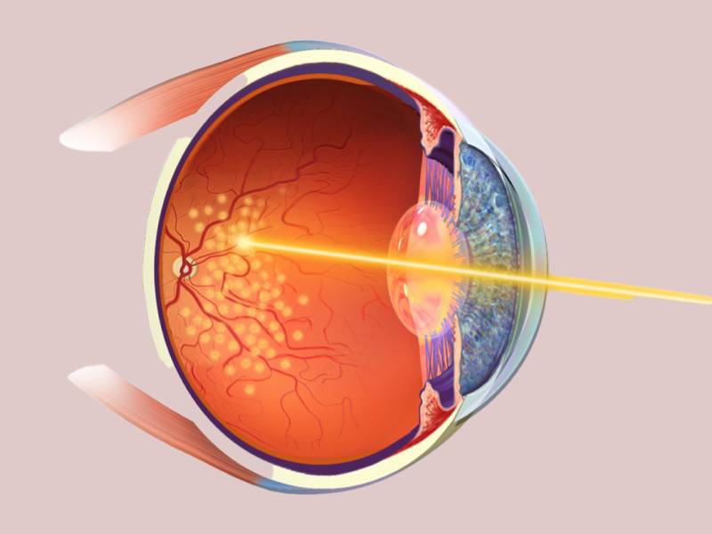
Imaging Tests for Detailed Analysis
Sometimes additional imaging tests are recommended to get more precise information about the state of your eyes. These include Optical Coherence Tomography (OCT), which uses light waves to create high-resolution cross-sectional images of retinal layers. Fluorescein angiography may also be employed if there’s suspicion that blood flow issues are contributing factors; this test involves injecting a dye into your bloodstream while taking photos of your retinal vessels.
Differentiating Retinal Detachment From Other Conditions
Some symptoms associated with retinal detachment overlap with other conditions like posterior vitreous detachment (PVD) or macular degeneration. The diagnostic process focuses on distinguishing these issues from true retinal detachment since their treatments differ significantly. For example, PVD often resolves on its own without intervention, whereas untreated retinal detachment can lead to irreversible vision loss.
When Should You See a Specialist?
If you experience sudden changes in vision such as flashes, floaters that increase in number rapidly, or partial vision loss described as "curtaining," seeking immediate medical attention is essential. Delaying diagnosis risks worsening damage and complicates treatment outcomes.
Accurate diagnosis requires both skillful examination by an eye specialist and sophisticated diagnostic equipment. By understanding these steps and actively monitoring potential warning signs in your daily life, you can ensure timely medical care if needed—ultimately protecting one of your most vital senses from lasting harm.
Treatment Options and Surgical Procedures
When it comes to retinal detachment, swift and appropriate treatment is crucial to preserving vision. Advances in medical technology have made it possible to effectively address retinal detachment through various treatment options, which are determined based on the severity, location, and underlying cause of the condition. Understanding these treatment modalities can empower patients with knowledge about what to expect during their care journey.
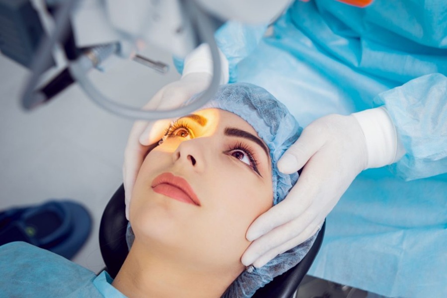
Laser Photocoagulation
One of the most common procedures for treating a small tear or hole in the retina is laser photocoagulation. During this procedure, a focused laser beam is used to create tiny burns around the edges of the retinal tear. These burns help seal the retina against the underlying tissue (the choroid), preventing fluid from seeping underneath and causing further detachment. This approach is typically performed on an outpatient basis, requires no surgical incision, and has a relatively short recovery time. Patients may experience some mild discomfort after the procedure but can usually return to their normal activities within a few days.
Cryopexy
Cryopexy is another technique used to treat certain types of retinal tears or detachments. This procedure is often used for peripheral tears that have caused minimal detachment but pose a risk of progression if left untreated. Like laser photocoagulation, cryopexy can often be performed in an outpatient setting.
Pneumatic Retinopexy
For more significant cases of retinal detachment, pneumatic retinopexy might be recommended. The gas bubble presses against the detached retina and holds it in position while scar tissue forms to seal any tears or holes. Patients undergoing this treatment may need to maintain specific head positions for several days or weeks to ensure proper healing.
Scleral Buckling
In cases where larger areas of retinal detachment are present, scleral buckling may be recommended as a surgical option. This reduces traction on the retina and allows it to reattach effectively. Scleral buckling has been successfully used for decades and remains one of the most reliable methods for treating extensive detachments.
Vitrectomy Surgery
Vitrectomy is another advanced surgical option commonly employed when other procedures are insufficient or when complications arise due to scarring or severe detachment involving central vision structures like the macula. In this procedure, tiny instruments are used to remove gel-like vitreous fluid from inside the eye along with any debris contributing to traction on the retina. Once cleared, either gas or silicone oil may be injected into its place temporarily until reattachment occurs successfully.
Post-Treatment Considerations
Regardless of which treatment option is pursued, follow-up care plays an essential role in ensuring long-term success and preventing recurrent issues. Many patients will require regular monitoring by their ophthalmologist after treatment so that healing progress can be tracked closely.
Ultimately, timely intervention combined with adherence to post-treatment recommendations gives individuals affected by retinal detachment an excellent chance at preserving their vision while minimizing future risks associated with this condition.
Tips for Protecting Your Vision and Reducing Risk
Protecting your vision and reducing the risk of retinal detachment is crucial for maintaining long-term eye health. While some factors, such as age or genetics, cannot be controlled, there are several proactive measures you can take to reduce your risk and safeguard your eyesight.
Prioritize Regular Eye Exams
One of the most effective ways to protect your vision is by scheduling comprehensive eye exams regularly. These exams allow eye care specialists to detect early signs of retinal issues or other underlying conditions that could increase the likelihood of detachment. For individuals with high-risk factors—such as a family history of retinal disorders, high myopia, or previous eye surgeries—more frequent screenings may be recommended.
Manage Chronic Health Conditions
Chronic health issues like diabetes and hypertension can significantly impact eye health. Uncontrolled blood sugar levels can damage the small blood vessels in the retina, increasing the risk of retinal detachment or other complications like diabetic retinopathy. Similarly, elevated blood pressure can lead to increased strain on delicate retinal tissues. Working with healthcare providers to manage these conditions effectively reduces overall risk.
Protect Your Eyes from Trauma
Blunt force trauma or injury to the eyes is a major cause of retinal detachment in many cases. Wearing appropriate protective gear during sports or when working with tools and machinery is essential to minimize this risk. For active individuals, safety goggles provide an extra layer of defense against accidental impacts that could harm ocular structures.
Be Mindful of Sudden Vision Changes
Being attuned to any sudden changes in your vision is an important part of reducing risks associated with retinal detachment. Flashes of light, a sudden increase in floaters, or shadows appearing in peripheral vision should never be ignored. Seeking immediate medical attention when these symptoms arise ensures timely intervention before significant damage occurs.
Avoid High-Risk Activities When Possible
Certain activities—such as skydiving, scuba diving at extreme depths, or high-impact sports—can sometimes increase intraocular pressure (IOP) or create risks for physical trauma that might lead to retinal complications. Consulting an ophthalmologist about safe practices related to specific hobbies is advisable if you have predisposing factors for retinal detachment.
Maintain a Healthy Lifestyle
A well-rounded diet that includes nutrient-rich foods supports better overall eye health. Foods high in antioxidants—such as leafy greens (spinach, kale), carrots (rich in beta carotene), oranges (vitamin C), and fish rich in omega-3 fatty acids—help maintain strong retinal cells and reduce oxidative stress on ocular tissues. Staying hydrated also ensures proper circulation within the eyes.
Regular exercise further contributes by improving blood flow throughout the body, including to the retina—reducing risks associated with poor circulation while also lowering systemic conditions like diabetes and hypertension that can harm vision over time.
Know Your Risks Based on Eye History
If you’ve undergone previous surgeries such as cataract removal or laser treatments involving your eyes—or if you’ve had prior instances of posterior vitreous detachment—it’s crucial to stay vigilant about monitoring symptoms related to potential complications in those areas.
By adopting these preventative strategies into daily life and maintaining open communication with trusted healthcare providers, anyone can take meaningful steps toward safeguarding their visual health while significantly reducing their chances of developing problems like retinal detachment over time.
Recovery, Follow-Up Care, and Long-Term Outlook
Recovering from retinal detachment is a critical phase that requires a combination of medical care, lifestyle adjustments, and patience. After undergoing treatment—whether surgical or non-surgical—the eye needs time to heal, and proper follow-up care is essential to prevent complications or recurrence.
The Recovery Period
The recovery timeline can vary depending on the severity of the detachment and the type of treatment administered. For instance, patients who undergo surgery like scleral buckling or vitrectomy may require several weeks to months for full healing.
Positioning: If a gas or silicone bubble was used during surgery to help reattach the retina, patients are typically advised to maintain a specific head position. This ensures the bubble stays in contact with the affected area.
Avoiding Strain: Activities such as heavy lifting, bending over abruptly, or exercising vigorously should be avoided until cleared by an eye specialist.
Protecting the Eye: It's common for surgeons to recommend wearing an eye shield while sleeping to prevent accidental trauma. Additionally, sunglasses may be suggested during daytime hours for comfort and protection from light sensitivity.
Importance of Follow-Up Care
Regular follow-up visits with your ophthalmologist are crucial during recovery. These appointments allow your doctor to monitor the progress of healing, check for any signs of complications such as infection or elevated intraocular pressure, and assess how well your vision is restoring.
For some individuals, vision improvement can be gradual. While many experience stabilization within weeks after surgery, others may notice changes in vision over several months. It’s important to discuss any concerns with your doctor during these visits since early detection of new issues can lead to more effective interventions.
Addressing Long-Term Vision Changes
Even after successful recovery from retinal detachment, some patients may experience lasting visual changes such as reduced sharpness or difficulty perceiving depth. In cases where permanent damage has occurred due to delayed treatment or extensive detachment before surgery, low-vision aids like magnifiers or specialized glasses can help improve quality of life.
Maintain annual eye exams even if you feel fine.
Monitor for symptoms in either eye that might suggest a new problem since individuals who've had one retinal detachment have an increased risk for it happening again in their other eye.
Emotional Adjustment and Support
Coping with vision loss—or even temporary impairment—can have emotional impacts that shouldn’t be overlooked. Feelings of anxiety about recurrence are normal but manageable with reassurance from your healthcare team and support groups tailored for those living with retinal issues.
Creating healthy habits not only aids physical recovery but also helps foster mental well-being over time. By staying proactive about follow-up care and maintaining open communication with your medical team regarding any concerns post-treatment, you’ll be better equipped to adapt to any long-term outcomes while preserving overall visual health.


© 2026 Memorial Vision - All Rights Reserved - Privacy Policy - Accessibility Statement
Powered by